


A fracture is a condition in which there is a break in the continuity of the bone. In younger individuals, these fractures are caused by high-energy injuries, as from a motor vehicle accident. In older people, the most common cause is weak and fragile bones.
Fractures of the knee can include the following:
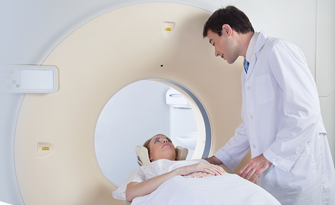
Diagnosis is made through your medical history, physical examination and other diagnostic imaging tests. X-rays are taken to determine whether the bone is intact or broken. X-rays are also helpful to know the type and location of the fracture. Your doctor may also recommend a computerized tomography (CT) scan to determine the severity of the fracture.
Treatment options include non-surgical and surgical methods. Non-surgical treatment options involve skeletal traction, and use of casts and braces. Skeletal traction involves placement of a pin into the bone in order to realign the broken bones. Surgery involves internal fixation and external fixation.
Internal fixation may involve:
During the procedure, metal pins or screws are inserted into the middle of the femur and tibia, and are attached to a device outside the skin to hold the bone fragments in place, and to allow alignment and healing.
If your bone is fractured in many pieces, a plate or rod is fixed at both ends of the fracture to maintain the overall shape and length of the bone while it heals. In elder patients where fracture healing delays, a bone graft taken from the patient or tissue bank may be used to form callus. In severe cases, the bone fragments are removed and the bone is replaced with a knee replacement implant.
The most common complications of surgery include infection, knee stiffness, delayed bone healing and knee arthritis.