


Patellofemoral instability is the moving out of your patella or kneecap from its normal alignment in the knee.
Patellofemoral instability can be caused because of variations in the shape of the patella or its trochlear groove as the knee bends and straightens. Normally, the patella moves up and down within the trochlear groove when the knee is bent or straightened. Patellofemoral instability occurs when the patella moves either partially (subluxation) or completely (dislocation) out of the trochlear groove.
A combination of factors can cause this abnormal tracking:
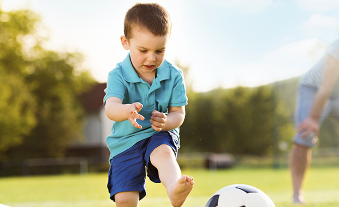
Young active individuals involved in sports activities are more prone to patellofemoral instability.
Patellofemoral instability causes pain when standing up from a sitting position and a feeling that the knee may buckle or give way. When the kneecap slips partially or completely, you may have severe pain, swelling, bruising, visible deformity and loss of function of the knee. You may also have changes in sensations such as numbness or even partial paralysis below the dislocation as a result of pressure on the nerves and blood vessels.
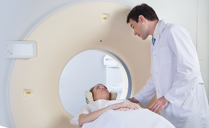
Your doctor evaluates the source of patellofemoral instability based on your medical history and physical examination. Other diagnostic tests, such as X-rays, MRI and CT scan, may be ordered to determine the cause of your knee pain and rule out other conditions.
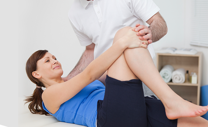
If your kneecap is only partially dislocated (subluxation), your physician may recommend nonsurgical treatments, such as pain medications, rest, ice, physical therapy, knee-bracing and orthotics. If the kneecap has been completely dislocated, it may need to be repositioned back in its proper place in the groove. This process is called closed reduction.
Surgery is sometimes needed to help return the patella to a normal tracking path when other nonsurgical treatments have failed. The aim of surgery is to realign the kneecap in the groove and decrease the Q angle.
Patellar realignment surgery is broadly classified into proximal re-alignment procedures and distal re-alignment procedures.
Proximal re-alignment procedures: During this procedure, structures that limit the movements on the outside of the patella are lengthened or ligaments on the inside of the patella are shortened.
Distal re-alignment procedures: During this procedure, the Q angle is decreased by moving the tibial tubercle towards the inner side of the knee.
The surgery is performed under sterile conditions in the operating room, under spinal or general anesthesia. The surgeon makes two or three small cuts around your knee. The arthroscope, a narrow tube with a tiny camera on the end, is inserted through one of the incisions to view the knee joint. Specialized instruments are inserted into the joint through other small incisions. The camera attached to the arthroscope displays the image of the joint on the monitor. A sterile solution is pumped into your knee in order to stretch the knee and provide a clear view and room for the surgeon to work. With the images from the arthroscope as a guide, the surgeon looks for any pathology or anomaly, and repairs it through the other incisions using various instruments. After the evaluation is completed, a larger incision is made over the front of the knee. Depending on your situation, a lateral retinacular release may be performed. In this procedure, the tight ligaments on the outer side of the knee are released, thus allowing the patella to sit properly in the femoral groove. Your surgeon may also tighten the tendons on the inside, or medial side of the knee to realign the quadriceps.
In cases where the malalignment is severe, a procedure called a tibial tubercle transfer (TTT) is performed. In this procedure, a section of bone where the patellar tendon attaches to the tibia is removed. This bony section is then shifted and properly realigned with the patella and reattached to the tibia using screws. Once the malalignment is repaired and confirmed with arthroscopic evaluation, the incisions are closed with sutures.
Your doctor will recommend medications to relieve pain. To help reduce swelling, you will be instructed to elevate the leg and apply ice packs to the knee. Crutches are necessary for the first few weeks to prevent weight-bearing on the knee. A knee immobilizer may be used to stabilize the knee. You will be instructed about the activities to be avoided and exercises to be performed for a faster recovery. A rehabilitation program may be advised for a speedy recovery.
Possible risks and complications associated with the surgery include:
Patients with patellofemoral instability have problems with the alignment of the kneecap. Therefore, treatment is necessary to bring the kneecap back into normal alignment. Your surgeon will decide which procedure is appropriate for your particular situation.