


The femur or thighbone is the longest and strongest bone in the body, connecting the hip to the knee. A femur fracture is a break in the femur. Depending on the location of the fracture, thighbone fractures can be categorized into:
Femur fractures may be caused by high-energy injuries such as a fall from a height or a motor vehicle accident. Patients with osteoporosis, bone tumor or infections, or a history of knee replacement are more prone to femur fractures. In the elderly, even a simple fall from a standing position may result in a fracture as the bones tend to become weak and fragile with advancing age.
Sudden, severe pain, along with swelling and bruising, are the predominant symptoms of femur fracture. The site is tender to touch with a visible physical deformity and shortening of the leg.
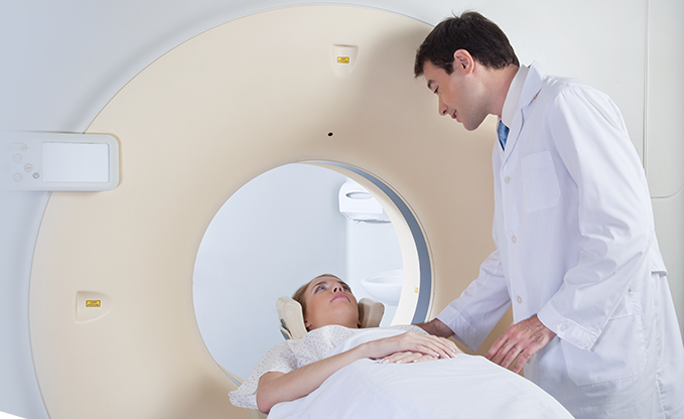
The diagnosis of femur fracture is based on the patient’s medical history, including the history of any previous injury, complete physical examination and imaging studies. The doctor will evaluate the soft tissue around the joint to identify any signs of nerve or blood vessel injury. Multiple X-rays and other imaging studies, such as CT and MRI scans, may be ordered to identify the location and severity of the fracture.
The management of the fracture is based on the severity of the fracture, medical condition of the patient and the patient’s lifestyle. It can be done by non-surgical or surgical methods.
Non-surgical treatment comprises of immobilizing the fracture site with the help of casts or braces to prevent weight-bearing and help in the healing process. X-rays are taken at regular intervals to assess the healing process. Weight-bearing and movement are initiated gradually, depending on the nature of the injury and the condition of the patient.
Surgical treatment is considered to realign the fractured bone. The use of advanced technology and special materials have improved the surgical outcome even in older patients. External or internal fixation or a knee replacement may be required, depending on the extent of the fracture.
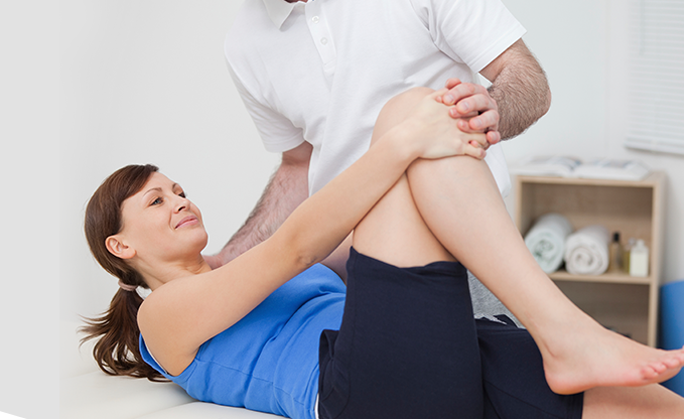
Rehabilitation of the femur fracture depends upon several factors such as age, general health of the patient and the type of fracture. As the femur fracture usually involves the weight-bearing joint, it may cause long-term problems such as loss of knee motion or instability, and long-term arthritis. Hence, a rehabilitation program is initiated along with the treatment, which comprises of instructions on weight-bearing, knee movements and the use of external devices such as braces.