Fractures/Dislocations

A bone fracture is a medical condition in which a bone is cracked or broken. It is a break in the continuity of the bone. A bone may get fractured completely or partially, and is commonly caused from trauma due to a fall, motor vehicle accident or sports injuries. Thinning of the bone due to osteoporosis in the elderly can also cause the bone to break easily. Overuse injuries are common causes of stress fractures in athletes.

Types of fractures include:

  • Simple fractures, in which the fractured pieces of bone are well aligned and stable.
  • Unstable fractures are those in which fragments of the broken bone are misaligned and displaced.
  • Open (compound) fractures are severe fractures in which the broken bones cut through the skin. This type of fracture is more prone to infection and requires immediate medical attention.
  • Greenstick fractures are unique fractures that occur in children. It involves the bending of the bone without any breakage.

Fracture healing

Our body reacts to a fracture by protecting the injured area with a blood clot, and callus or fibrous tissue. Bone cells begin forming on either side of the fracture line. These cells grow towards each other and thus close the fracture.

Medical therapy

The objective of early fracture management is to control bleeding, prevent ischemic injury (bone death) and remove sources of infection, such as foreign bodies and dead tissue. The next step in fracture management is the reduction of the fracture and its maintenance. It is important to ensure that the involved part of the body returns to its function after the fracture heals. To achieve this, maintenance of the fracture reduction with immobilization technique is done by either non-operative or surgical methods.

Non-operative (closed) therapy 

Non-operative (closed) therapy comprises of:

  • Casting closed reduction is done for any fracture that is displaced, shortened or angulated. Splints and casts made up of fiberglass or plaster of Paris material is used to immobilize the limb.
  • Traction method is used for the management of fractures and dislocations that cannot be treated by casting. There are two methods of traction namely, skin traction and skeletal traction.
    • Skin traction involves attachment of traction tapes to the skin of the limb segment below the fracture.
    • In skeletal traction, a pin is inserted through the bone distal to the fracture. Weights will be applied to this pin, and the patient is placed in an apparatus that facilitates traction. This method is most commonly used for fractures of the thighbone.

Surgical therapy

  • Open reduction and internal fixation (ORIF): This is a surgical procedure in which the fracture site is adequately exposed and the fracture is reduced. Internal fixation is done with devices such as Kirschner wires, plates and screws, and intramedullary nails.
  • External fixation: External fixation is a procedure in which the fracture stabilization is done at a distance from the site of fracture. It helps to maintain bone length and alignment without casting.

External fixation is performed in the following conditions:

  • Open fractures with soft-tissue involvement
  • Burns and soft tissue injuries
  • Pelvic fractures
  • Comminuted and unstable fractures
  • Fractures having bony deficits
  • Limb-lengthening procedures
  • Fractures with infection or non-union

Rehabilitation

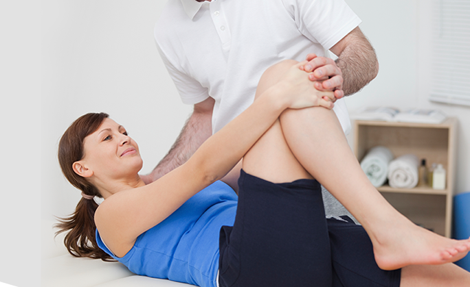
Fractures may take several weeks to months to heal completely. You should limit your activities even after the removal of the cast or brace so that the bone becomes solid enough to bear the stress. Rehabilitation program involves exercises and gradual increase in activity levels until the process of healing is complete.

Foot and Ankle

The foot and ankle in the human body work together to provide balance, stability, movement and propulsion.

This complex anatomy consists of:

  • 26 bones
  • 33 joints
  • Muscles
  • Tendons
  • Ligaments
  • Blood vessels, nerves and soft tissues

Hip

The thigh bone (femur) and the pelvis (acetabulum) join to form the hip joint. The hip joint is a “ball and socket” joint. The “ball” is the head of the femur and the “socket” is the cup shaped acetabulum. The joint surface is covered by a smooth articular surface called cartilage that cushions the joint and allows pain-free movement. This cartilage does not show up on X-ray, therefore you can see a “joint space” between the femoral head and acetabular socket.

Leg

Shoulder, Arm, Elbow